![An Australian Government Initiative [logo]](/images/austgovt_brown_90px.gif)

Two Groups - classifying fungi into ascomycetes and basidiomycetes:
Truffle-like fungi - basidiomycetes
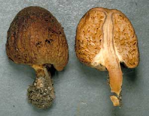
 Setchelliogaster sp. (right) |
 |
The fruiting bodies of the basidiomycete truffle-like fungi are varied in form,
sometimes stalked but mostly stalk-less and more-or-less spherical in shape
![]() .
Internally the fruiting bodies are chambered, with the chambers of some species
easy to see with the naked eye - but a hand lens is needed to see the individual
chambers in other species. The basidia line the walls of the chambers and protrude
into the interiors of the chambers - which are empty in many species, but not
in all. In shape the chambers may be anything from spherical to quite contorted.
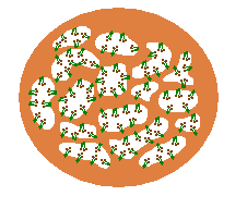
This diagram represents the cross-section of a truffle-like fruiting body. The
solid, fleshy areas of the truffle are coloured brown and you can see the basidia
(coloured green and bearing dark brown spores) lining the walls of those chambers.
Of course, this diagram exaggerates the sizes of both the basidia and the chambers
and this simplistic two-dimensional figure does not do justice to the internal
convolutions of the three-dimensional fruiting body. The photograph of the stalked
Setchelliogaster fruiting body may give you a better idea of the internal
structure. On the left you can see the outside appearance and on the right the
internal structure. In the latter you can see some of the chambers (of varied
forms) and get an idea of the intricate way in which the internal tissue creates
those chambers.
.
Internally the fruiting bodies are chambered, with the chambers of some species
easy to see with the naked eye - but a hand lens is needed to see the individual
chambers in other species. The basidia line the walls of the chambers and protrude
into the interiors of the chambers - which are empty in many species, but not
in all. In shape the chambers may be anything from spherical to quite contorted.
This diagram represents the cross-section of a truffle-like fruiting body. The
solid, fleshy areas of the truffle are coloured brown and you can see the basidia
(coloured green and bearing dark brown spores) lining the walls of those chambers.
Of course, this diagram exaggerates the sizes of both the basidia and the chambers
and this simplistic two-dimensional figure does not do justice to the internal
convolutions of the three-dimensional fruiting body. The photograph of the stalked
Setchelliogaster fruiting body may give you a better idea of the internal
structure. On the left you can see the outside appearance and on the right the
internal structure. In the latter you can see some of the chambers (of varied
forms) and get an idea of the intricate way in which the internal tissue creates
those chambers.