![An Australian Government Initiative [logo]](/images/austgovt_brown_90px.gif)

Two Groups - classifying fungi into ascomycetes and basidiomycetes:
Flask fungi
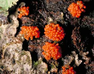 Nectria fruiting bodies |
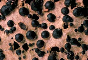 Perithecia on dead wood |
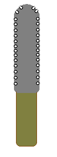
The Flask Fungi produce their spores in tiny, generally spherical, chambers
(called perithecia) which are usually under a couple of millimetres in
diameter. The first photo shows a number of tiny orange balls, each only about
a millimetre or two in diameter. Each such ball is a separate perithecium and
is the fruiting body of a species of Nectria. Here's another photo showing
some black hemispheres (about 2mm in diameter) on a piece of dead wood. Once
again these are examples of perithecia. If you cross-sectioned a perithecium
you'd see the asci inside, as shown by this diagram.
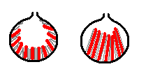
Of course, this is a very simplistic diagram and there is considerable variation in both the shapes and contents of perithecia. These diagrams show some of the variation in shapes that can be seen in different species.
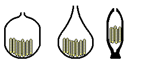
In some species the chamber may be relatively empty, others may have paraphyses or even have the entire chamber filled. The asci may be found only at the base of the perithecium - or up the sides as well. Two examples are shown here (with the asci in red and paraphyses in grey)
|
Diagram below |
|
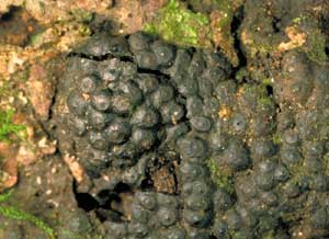
Just as there are "compound apothecial" fruiting bodies (such as Morchella and Cyttaria), so there are also "compound perithecial" fruiting bodies, where a number of perithecia are combined within a larger structure. In many perithecial ascomycetes the perithecia are embedded within a communal tissue (called a stroma, with the plural stromata). In many cases the stroma forms a dark-coloured, brittle sheet on wood as shown in this photo of Hypoxylon sp. The little pimples that dot the black sheet are the very tips of the perithecia. In cross-section such a compound fruiting body would have the following structure.The mass of dark grey represents the stroma in which the perithecia are embedded and you can see that the apical openings just protrude above the general level of the stroma.
 |
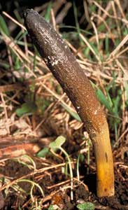 Cordyceps gunnii |
As well as such sheet-like species there are various species with markedly
three-dimensional stromata. The species of Cordyceps are parasites, mostly
of invertebrates and commonly of invertebrate larvae. The fruiting body in this
photo (of Cordyceps gunnii, ) has emerged from such a parasitized larva
in the ground ![]() .
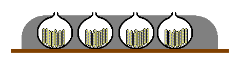
In cross-section the fruiting body would resemble the following diagram, with
the perithecia just under the surface of the stroma (again shown in grey). The
sterile stem is shown in khaki-green. Several species of Xylaria
.
In cross-section the fruiting body would resemble the following diagram, with
the perithecia just under the surface of the stroma (again shown in grey). The
sterile stem is shown in khaki-green. Several species of Xylaria ![]() have a similar club-like appearance and internal structure (though without the
well-differentiated stem of Cordyceps). However, while these club-like
fruiting bodies are of similar shape to those of genera such as Geoglossum
have a similar club-like appearance and internal structure (though without the
well-differentiated stem of Cordyceps). However, while these club-like
fruiting bodies are of similar shape to those of genera such as Geoglossum
![]() and Trichoglossum
and Trichoglossum ![]() (and Xylaria is even black), the internal constructions are quite different.
Remember that Geoglossum and Trichoglossum have no perithecia
but have a palisade of asci and paraphyses lining their surfaces. This is explained
in the CUP FUNGI section.
(and Xylaria is even black), the internal constructions are quite different.
Remember that Geoglossum and Trichoglossum have no perithecia
but have a palisade of asci and paraphyses lining their surfaces. This is explained
in the CUP FUNGI section.
 Daldinia concentrica (left) diagram (above) section through fruiting body (right) |
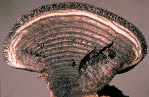 |
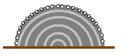
Another example of a perithecial ascomycete with a three-dimensional stroma
is Daldinia concentrica ![]() ,
which produces hemispherical fruiting bodies (up to several centimetres in diameter)
on dead wood. One photo shows a cross-section, which is also illustrated in
this diagram - with the perithecia around the periphery.
,
which produces hemispherical fruiting bodies (up to several centimetres in diameter)
on dead wood. One photo shows a cross-section, which is also illustrated in
this diagram - with the perithecia around the periphery.
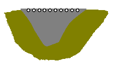 |
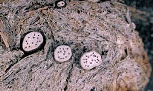 Poronia erici growing on dung |
You may often see small, black-spotted, whitish disks on herbivore dung, such
as shown in this photo. These are the fruiting bodies of a perithecial ascomycete
called Poronia erici and may grow to about a centimetre across. The black
dots are the tips of the embedded perithecia and a cross-section would show
a view much as the one in this diagram, with the perithecia under the upper
surface of the grey stroma. The khaki green represents the surrounding dung.